BCA staining test

Quantification of cellular protein content
Measurement of total cellular protein as an indicator of cell growth and viability.
The BCA staining test is an in vitro cytotoxicity assay based on the bicinchoninic acid (BCA) method for the determination of cellular protein content.
The BCA reagents consist of a water-soluble and stable BCA (bicinchoninic acid) solution and an alkaline Cu²⁺ solution.
The amino acids cysteine, cystine, tryptophan and tyrosine, which are constituents of cellular proteins, bind to these reagents and reduce Cu²⁺ to Cu⁺. The resulting Cu⁺ ions react with bicinchoninic acid to form a water-soluble violet dye.
The intensity of the violet colour correlates with the number of cells present in the culture and is measured by absorbance.
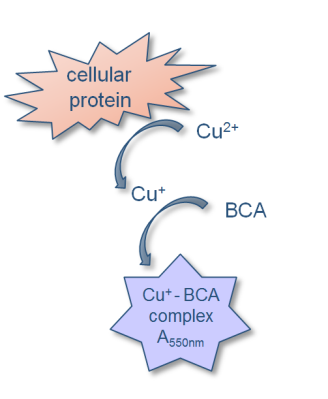
Assessment of growth inhibition of cells by extraction method and BCA staining
- The test item is extracted under agitation for a defined period in DMEM supplemented with 10% FBS at 37 ± 1°C, and L929 cells are incubated with the extract.
- Near the end of the incubation period, cells are evaluated microscopically.
- Following incubation, cells are washed with buffer and the BCA reagents are added. Cells are incubated with the reagents for 0.5 to 2 hours, depending on the absorbance measured at 550 nm.
- According to ISO 10993-5, cytotoxic effects can be evaluated based on the protein content of the cultures, which serves as a measure of cell growth.
- A growth inhibition of more than 30% in cultures treated with the test extract compared with untreated control cultures (solvent control) is considered a clear cytotoxic effect.
BCA staining test protocol
Protocol |
|
|
Cell line |
L929 cells (ATCC No. CCL1, NCTC clone 929; connective tissue mouse cell line, clone of strain L, DSMZ) |
|
Analysis |
The BCA assay consists of two reactions: 1. Protein peptide bonds reduce Cu2+ ions to Cu+ 2. Two bicinchoninic acid molecules chelate with a Cu+ ion, resulting in a violet complex that absorbs light at a wavelength of 550 nm |
|
Concentrations |
4 concentrations of the test extract: 29.6%, 44.4%, 66.7% and 100% |
|
Extraction time |
4 - 72 h at 37 ± 1°C - 4 h (short-term contact and intact skin or mucosa) |
|
Incubation time |
24 - 72 h at 37 ± 1°C |
|
Quality controls |
Solvent Control: DMEM 10% FBS Negative control: Polypropylene extracted in DMEM 10% FBS Positive control: Latex extracted in DMEM 10% FBS |
|
Data delivery |
Growth Inhibition of L929 cells is determined by measuring protein content at 550 nm absorbance. |
|
Positive prediction |
Growth inhibition greater than 30% in cultures treated with the test extract compared with untreated control cultures (solvent control) is considered a clear cytotoxic effect. |
References
- Smith, P.K., Krohn, R.I., Hermanson, A.K., Mallia, A.K., Gartner, F.H., Provenzano M.D., Fujimoto E.K., Goeke N.M. Olson B.J. and Klenk D.C: 1985, “Measurement of Protein Using Bicinchoninic Acid.“, Analytical Biochemistry 150, 76 – 85
- ISO 10993-5: 2009, “Biological evaluation of medical devices – Part 5: Tests for in vitro cytotoxicity“
- ISO 10993-12: 2012 “Biological evaluation of medical devices – Part 12: Sample preparation and reference materials”